

What is a PROTEIN?
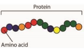
PROTEINS are large molecules playing important functions that make living things alive. They are made out of different combinations of 20 types of amino acids, like words out of a 20-letter alphabet — but long ones with hundreds of letters, even longer than 34,000 letters like the muscle protein titin! Each [ihc-hide-content ihc_mb_type=”show” ihc_mb_who=”2,3″ ihc_mb_template=”1″ ] sequence of amino acids will fold into a unique shape, which is crucial to how the protein will function.
The information on a protein’s sequence is stored in a gene, and our DNA contains at least 20,000 such genes. A set of the genes, known as housekeeping genes, code for proteins which are essential for keeping cells alive; every type of cell also turns on another set of genes coding for proteins specific for the cell’s functions. White blood cells make and secrete proteins called antibodies that fight infections, while connective tissue cells secrete collagen — the most plentiful protein, about a quarter of all the protein, in our body — that make up the skin and bones that hold our body together.
How are protein structures studied?
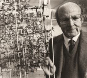
X-RAY CRYSTALLOGRAPHY, developed in the 1910s, was used to solve the first protein structures by Max Perutz ( pix ) in the late 1950s. The method involves turning a protein into a crystal — a process requires large quantities of purified protein as well as, as some scientists put it, dumb luck — then shooting an X-ray beam through it, and from the resulting diffraction pattern calculating the electron density of every atom in the crystal. Today, about 140,000 structures have been solved with this method.
Perutz, a young scientist then, started working on haemoglobin in 1937. He soon got beautiful X-ray diffraction patterns from his haemoglobin crystals, but at that time no one could solve the structure due to a mathematical issue, the phase problem, which became very complex for large molecules like proteins. It took him 16 years to make a breakthrough — soaking his crystals in mercury solutions and comparing the diffraction pattern against the one without mercury — and a further six years to improve the method and make the complicated calculations, finally determining the structure in 1959 after 22 years of hard work. Perutz was awarded a Nobel Prize in 1962; the lab that he set up produces in total 15 Nobel laureates, including Watson and Crick who cracked the DNA structure.
How do proteins function?
PROTEINS function by interacting with other molecules; the distinct shape, or structure, of a protein enables it to recognise and bind to specific molecules, roughly like lock and key. For example, titin binds to other muscle proteins and acts as a molecular spring — it unfolds when stretched and refolds when the force is removed, returning muscle to the proper shape.
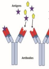
When rhodopsin in our eyes detects light, it changes shape and causes a signal to be sent to our brain. Antibodies, meanwhile, bind to foreign molecules, such as those on a bacterial surface, as markers for destruction by white blood cells; every time our body encounters a new foreign particle, it develops antibodies that bind to it tightly.
How does haemoglobin transport oxygen?
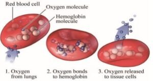
IN ADDITION to amino acids, haemoglobin in the red blood cells also contains a haem group with an iron atom, which makes our blood red.
Oxygen binds to the iron atom in our lungs, and is released in the rest of our body where the oxygen level drops. A change in the haem surroundings of a bacterial haemoglobin, HGbI, makes it bind oxygen 800 times more tightly than our haemoglobin — were HGbI used as artificial blood, we would suffocate as it would grab all the oxygen in our body!
What is an enzyme?
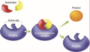
ENZYMES are proteins that, after binding to their target molecules, speed up reactions that either break the targets apart or combine them into new molecules — reactions which otherwise may happen million times more slowly by themselves.
Amylase in our saliva, for example, breaks down starch into sugar. Complete breakdown of glucose into CO2 and water involves another two dozen enzymes, and releases energy that is stored in ATP, the molecular currency of energy, whose breakdown in turn will provide energy to drive reactions like synthesis of proteins themselves. The enzyme ATP synthase (can you guess its function?), the world’s tiniest motor, has two rotary motors that rotate in opposite directions: every revolution the motors make three ATP molecules are generated. Glucose is also converted into another sugar, ribose-5phosphate which is used for DNA synthesis, in a process involving G6PD; a defect in this enzyme leads to G6PD deficiency and causes red blood cells to break down prematurely.
A recent exciting discovery is a bacterium that produces two plastic-degrading enzymes, which turn PET bottles into food for the bacterium; without these enzymes, the bottles may take up to 1,000 years to break down. Scientists have also identified enzymes that, on the other hand, combine certain molecules into polyhydroxyalkanoate (PHA), a type of biodegradable bioplastic which has the potential to replace conventional plastics, and which has been used in making absorbable medical devices such as sutures.
And why are they studied?
FOR SEVERAL good reasons — besides getting to know how haemoglobin binds oxygen, and how a change of an amino acid, or a mutation, causes it to stick to each other into fibres that distort red blood cells and lead to sickle cell disease. Studying protein structures helps scientists design better enzymes too.
The plastic-degrading enzyme, PETase, is exciting, but it doesn’t act fast enough to solve humans’ plastic waste woes; determining its structure has given scientists hints to come out with an improved version. Detergents are added with enzymes to help remove food stains, and their structures are enabling the laundry industry to engineer versions that can withstand high temperatures and highly alkaline conditions.
Knowing the structure of a protein also helps scientists design keys that can fit the lock — and cure a disease. The drug zanamivir was designed to bind to the enzyme neuraminidase of influenza viruses, rendering newly formed viruses unable to detach from infected cells. Armed with structural information, scientists developed imatinib to prevent Bcr–Abl tyrosine kinase, a fusion enzyme that causes a form of leukaemia, from acting on its targets, and the drug has been a phenomenal success in treating the cancer.